
Library Record
Images
Additional Images [1]
Metadata
Catalog Number |
2016.25.693 |
Accession number |
2016.25 |
Object Name |
Book |
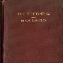
Title |
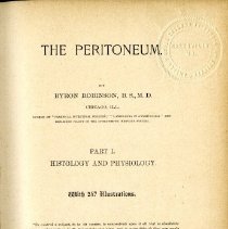
The Peritoneum Part I Histology and Physiology |
Published Date |
1897 |
Summary |
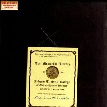
The Peritoneum Part I Histology and Physiology; 1897. Written by Byron Robinson, published by the W. T. Keener Company in Chicago, with 247 illustrations, maroon cover with gold lettering on front cover and spine, Memorial Library sticker attached to front interior cover "Mrs. Geo. M. Laughlin"; 17.8 cm (w) x 26.2 cm (h), 103 pages. Cover worn and faded, black paint on spine with call number written in white along with small yellow sticker, spine extremes worn, paper yellowed with acid burn around the edges, front endpaper and several front pages are loose in the book, "Property of KCOS Library" stamped around edges of the textblock, library outcards attached to interior back cover and back endpaper. |
Credit line |
Museum of Osteopathic Medicine |
People |
Aeby Affannasiew Allerow Arnold Arsellio Auerbach Bajard Bardon-Sanderson Bartholin Beck Berkely, Henry J. Bichat Binz Birdsall Bizzozero Bordeau Bottcher Bourgery Bowman Brechet Brecke Brinton Bruns Cetacea Chrzonszczewsky Chrzozozinsky Chyle Chyme Clark Cohnstein Conheim, Julius Cruikshank Cruveilhler Cuvier Cyon, V. Dbar Deckhuyzen Desault Dogiel Donders Dujardin Dybkowsky Ebers Erasistratus Eustachius Fick, A. Finkham Flemming Fohman Frey Frye Fuhrer Galenus Gaskell Gegenbaur Gerber Glisson Goluben Goodsir Grew Gunning Haeckel Hale Haller Hamberger Harvey Heindenhain Heitzmann Henle Herbst Hering Hermann Hertwig, Oscar Hewson His Hiswas Hoffman Hooke Hunter, John Hunter, William Hyrtl Hyrtle Inzani Joyliffe Jullien, L. Key Klein Koeliker Kolossow Krause Kuhne Kundrat Lacuna Langerhans Laughlin, Blanche Still Lauth Lawdowsky Lazarus-Barlow, W. S. Leathes, J. B. Lessing Leuwenhoek Leydig Lieberkuehn Lobstein Loret Ludwig Luschka Maffuci Magendic Malpighi Mascagni Meckel Merkel Metchnikoff Meyer, Joseph Miller, J. Mirbel Mohl Monroe Muller Muscatello Nesterowsky Nikolsky Notkins Nuck Oedmansson Orlow Pacinni Paladine Panizza Pavia Pequet Pflueger Pinel Poisonille Ponfick Ponyes Prochaska Ptolemy Purkinje Radjewsky Ranvier Recklinghausen Reichert Reitz Remak Remy Retzii Rindfleisch Robin Robinson, Byron Rudbeck Rudolph Ruysch Salzmann Sappey Schklarewsky Schleiden Schmidt-Lauterman Schultze Schwann Schweigger-Seidel Settala Sheldon Stricker Tadino Teichmann Thoma Tiedemann Tod Toldt Tourneau Treitz Tubby Turpin Turtle Valentine Vater Virchow Von Recklinghausea Wadd Waldeyer Waller Wegner, George Wesling |
Search Terms |
Abdominal serosa Absorption Absorption by lymphatic system Absorption by vital course Absorption filtration Absorption fluid Absorption gas Absorption imbibition Absorption intraabdominal pressure Absorption of peritoneal fluids Absorption of proteids of tissue Absorption of salt Absorption of serous fluid Absorption oil Absorption osmosis Absorption stomata Absorptive capacity Action of leucocytes in regard to blood vessels Adipose tissue Adler and Meltzer Adventitia Albumen Amphibia Amphibian lymph sacs Amputated limbs Anastomosis Anastomosis of endothelial cells Anat minute Aorta abdominal Aorta thoracic Apertures Appendica epiploicae Appendicitis Arrangement of the endothelia Arteries internal mammary Ascites Auerbach plexus Author experiments and observations on the peritoneum Axis cylinder Bands peritonitic Bands pleuritic Basement membrane Beal and Schultze Berlin blue injections of Billroth-Meissner plexus Bladder Blood capillaries Blood plasma Blood serum Blood serum of hare Blood serum of ox Blood serum of sheep Blood stream Blood vascular capillaries Blood vessels Blood vessels development of Blood vessels of the peritoneum Canal interendothelial Canal vertical Capillaries lymph or blood Capillaries non-valved Capillaries of diaphragm Capillaries of intestines and spleen Capillary lymphatic fields Capillary pressure Capillary tension Cell and its establishment Cell fat borders Cell fat spaces Cell fat use of in peritoneum Cells Cells branched Cells connective tissue Cells elastic tissue Cells endothelial Cells fat Cells fibrillar Cells granular Cells granular polyhedral nucleated Cells lymphoid Cells muscle Cells nerve Cells pigment Cells vacuolated Cells vascular Cells vital action of Cells wandering Cement substance Centrum tendendum Centrum tendineum is a bed of lymphatics Centrum tendineum of diaphragmatica peritoneum Chronology of the peritoneum Cilia Ciliated endothelia Cisterna lympahtica magna Cleft Coagulation Colotomy Conclusions in regard to peritoneal endothelia Conclusions in regard to the blood vessels Conclusions in regard to the diaphragm Conclusions in regard to the interendothelial space Conclusions of absorption paths Conclusions on peritoneal absorption Conclusions on technique of peritoneum Condition of the interendothelial space and substance its physiology Conditions for the passage of leucocytes Connective tissue corpuscles Contents of the subperitoneal tissue Cords Corpuscle connective tissue Corpuscle lymph Cortical Cover plate Demonstration of lymphatics of diaphragm Diaphragm Diaphragm absorption of Diaphragm activity of Diaphragm and centrum tendineum its layers its peritoneal serosa Diaphragm experiment on Diaphragm is the special locality of the absorption of solid particles Diaphragm locality of absorption Diaphragm nerve supply Diaphragm steam toward Diaphragmatic lymphatics Diaphragmatic serosa Drainage Dropsical conditions of the peritoneum Dubar and Remy Ductus aguosi Eberth Edema Elastic fiber Elastic fibres Endothelia Endothelia blood vascular Endothelia diameter Endothelia germinating Endothelia grouping Endothelia growth of Endothelia lineaments Endothelia lymph vascular Endothelia nucleus Endothelia of blood vessels Endothelia of the free peritoneal surface Endothelia peritoneal Endothelia shape Endothelia spindle shaped Endothelial arrangement of Endothelial cell Endothelial contraction and expansion Endothelial membrane Endothelial plate Endothelial tube Epiblast Epidermis Epithelia Essential coats of vessels Experiments of Adler and Meltzer Experiments of author Experiments of Starling and Tubby Experiments on animals Experiments on cat Experiments on dog Experiments on guinea pig Experiments on rabbits Fallopian tubes Fat Fat globules Fatty tissue Fibers Fibers elastic Fibers muscular Filtrate hypothesis Filtration Fine structure of peritoneum Fixed connective tissue cells Fluid Fluid ascitic Fluid colored Fluid dropsical Fluids extravascular Fluids intravascular Foa Follicles Friction of serous membranes Friction pressure From nann Function of endothelia Function of interstitial spaces Function of peritoneal Function vascular and lymph Functions of the stomata vera Ganglia Germinating endothelia Germinating patches and tracts Glands lumbar Glands mammary Glands mediastinal Glands pyloric Glands salivary Glands sweat Glands testicular Glisson capsule Gold chloride Gold chloride melron Granules colored Ground substance of the peritoneum Heliopolis Hernia Histology and physiology of the peritoneum Humidity Hypoblast Imbibition Imbibition capillary Imbibition molecular Imbibtion Infection Inflammation Influence of blood vessels on endothelia Injection Interendothelial line Interendothelial space Interendothelial tendinous spaces Interstitial space Interstitial space subperitoneal Interstitial spaces Intestinal mucosa Intestines small Intra-abdominal pressure Irregular elements Jugular vein Juice canals Karyokinesis Kidneys Kinds of animals used for experiments Kinds of cells Kinds of tissue and cells Kitt substance Lacteals Lamina inferior Lamina superficialis Lamination Layers of centrum tendineum Leipzig Physiologic Institute Leucocytes Leucocytes function of Leucocytes migration of Ligamenta lata Ligamentum latum Ligamentum peritoneal Ligation of innominate veins Ligation of lymphatics Ligation of thoracic ducts Line interendothelial Line of lymph endothelium Liver Lymph Lymph capillaries Lymph channels Lymph cleft Lymph corpuscles Lymph fluid Lymph gland Lymph hearts Lymph nodes Lymph sacs Lymph sinus Lymph spaces Lymph stream Lymph trunks Lymph tubes and the blood tubes have a common ground in the lymph spaces Lymph vessels Lymphangial tracts Lymphatic pathways Lymphatics of the diaphragm Lymphatics of the peritoneum Lymphatics origin of Lymphatics system Lymphoid cells Malter colored Medullary sheath Medullary tubes Membrana limitans Membrana mesenteria propria Membrane basement Membrane germinal Membrane serous Mesenchyma Mesenteries are not merely duplicatures of the peritoneum but primordial structures Mesenterium not perforated Mesentery Mesoblast Mesoblastic origin of the peritoneum Mesogaster frog Mesothelium Methods of absorption Methods of origin of peritoneum Methods of staining and study of the endothelia Methods of the preparations of specimens of the peritoneum for microscopical examinations Methods to discover the lymphatics Microscopical exam Nerve axial fibers Nerve endings Nerve network Nerve pain Nerve periphery Nerve peritoneal network Nerve supply Nerves Nerves medullated Nerves nerve cells Nerves nonmedullated Nerves of the peritoneum Nerves of the peritoneum medullated Nerves of the peritoneum methods of preparation Nerves of the peritoneum non-medullated Nerves of the peritoneum remak band Nerves of the peritoneum vater-pacinian corpuscle Nerves of the various parts of the peritoneum and of different animals Nerves of the visceral and parietal peritoneum Nerves peritoneal Nerves Remak nerve fibers Nerves Vater-Pacinian corpuscles Neurolemma Nodes of Ranvier Nodules Non-valved capillaries Nucleus Nutrition Omentum Origin of serous cavity by pressure Osmosis Osmosis of glucose Outlines of peritoneal endothelia Ova sac Pacinni corpuscle Pancreas Paraschites Parts of an endothelial cell Patches Paths of absorption of peritoneal fluid Paths of interstitial fluids Paths of peritoneal absorption Perforation Perforations in the membrana limitans Perforations in the membrana limitans of the diaphragm Pericardial cavity Perilymphangial Peritoneal absorption Peritoneal absorption by mechanical pressure Peritoneal bed Peritoneal cavity Peritoneal current Peritoneal fluid Peritoneal membrane Peritoneal plates Peritoneal serosa Peritoneum Peritoneum absorption Peritoneum cavity Peritoneum contraction distention Peritoneum currents Peritoneum endothelia Peritoneum fluid composition Peritoneum friction Peritoneum function Peritoneum ground substance Peritoneum has distinct structure and function and sensation Peritoneum histology Peritoneum histology and physiology of Peritoneum historical sketch Peritoneum humidity Peritoneum irritation Peritoneum joint cavity Peritoneum lymph or interstitial space Peritoneum membrana limitans Peritoneum methods of absorption Peritoneum motion Peritoneum nerves of Peritoneum physiology Peritoneum physiology of Peritoneum polish Peritoneum preparation Peritoneum preservation of Peritoneum rabbits Peritonitis Perivascular Perivascular space Physiology Physiology of the peritoneum Pia Pia foa Pica-duodeno-jejunalis Pig embryo Pigment cells of the peritoneum Pillars of Uskow Planes of tissue Plasmatic vascular tubes Plates stomatal Plexiform Plexus pampiniformis Polish Pores Potassium ferrocyanide Precipitate Preservation of the peritoneum Pressure capillary peritoneal Pressure mechanical Processes Proliferation Protoplasm Protoplasm reproduction centers Protoplasm retraction Ptosis Puerperal fever Puerperal peritonitis Puncture method Pus Radiation Reagents acetic acid Reagents acid chromic Reagents auric chloride Reagents berlin blue Reagents chloride of platinum Reagents eosin acid fuchsin Reagents formaline Reagents logwood Reagents Muller fluid Reagents osmic acid Reagents silver nitrate Reagents tannin Regeneration of endothelia Retroperitoneal abscess Retrosternal glands Rings Sacculation Salvioli Sarcode Sensation Sepsis Septic Septum transversum Serosa peritoneal Serosa pleural Serosity Shape of endothelia Shape of the endothelia Sheath subperitoneal Significance of the peritoneum Silver nitrate Silver nitrate staining Similarity of animals peritoneum in structure and function Solutions hypertonic Solutions isotonic Source of germinating cells Space interendothelial Space perivascular Spaces intercapillary Spaces interendinous Spaces interendothelial Spaces interstitial Spaces intravascular Spleen Starling Starling and Tubby Starling experiments Statements of thoma Stigmata Stomata Stomata accidents from reagents Stomata anatomical structure of Stomata diaphragmatic Stomata generative centers Stomata interendothelial Stomata of blood vessels Stomata regulate peritoneal currents Stomata spuria Stomata vera Stomata vera and spuria Stomata vera functions of Strands fibroelastic Study of the endothelial membrane Subbotin Subjects for discussion Subperitoneal tissue Subserous lymphatic vessels Substance endolyphangial Substance interendothelial Sweat Sympathetic nerves Table of experiments Tears Technique of peritoneum Tendinous bundles Tendon Term endothelia Thoracic cavity Thoracic duct Thoracic duct rabbits Three modes of origin of lymphatics Tissue adipose Tissue areolar Tissue connective Tissue corpuscles fixed Tissue corpuscles wandering Tissue elastic Tissue fatty Tissue fibrous reticulated Tissue mesoblastic Tissue spaces Tissue subperitoneal Tissue subserous Tissue white fibrous Trachea Tract alimentary Tract respiratory Trauma Tube Tunica adventitia Tunica intina Tunica media Tunica vaginalis Two kinds of medullated nerves Unsettled points Vacuolation Valved capillaries Valved lymphatic channels Valves lymph Valves on lymph trunks Vasa efferentia Vasa inferential Vater-Pacinian corpuscle Veins innominate Veins omphalomesenteric Veins subclavian Veins umbilical View of his Vital cell processes Vital processes Vivisection Walls Walls lymph channel Wharton jelly White fibres Zarvilski experiments |